Drug Reactions
- related: Dermatology
- tags: #dermatology
Drug reactions are an important consideration when weighing a differential diagnosis for any rash, as medications can produce almost any pattern of skin disease. The most common types of cutaneous adverse reactions are exanthematous/morbilliform, urticarial, fixed drug, photosensitivity, hypersensitivity vasculitis, and the severe cutaneous adverse reactions (see Dermatologic Emergencies). Medications can also induce an array of specific diseases including psoriasis, bullous pemphigoid, and cutaneous lupus erythematosus (Table 7). Discontinuing the causative drug is the first step in treating drug reactions.
A morbilliform (measles-like) or exanthematous drug eruption consists of symmetrically arranged erythematous macules and papules, some discrete and others confluent. Morbilliform reactions are the most common drug reactions and are most likely a type IV delayed hypersensitivity reaction. As such, the rash appears in the first or second week after drug exposure, although subsequent exposures can produce a reaction much more quickly. Can have eosinophillia and leukocytosis but no other systemic involvement. Patients develop pink papules and macules that coalesce symmetrically to form plaques. The papules are often dense and monomorphic and are accompanied by varying degrees of pruritus beginning on the trunk and progressing distally across the limbs (Figure 23). Palms and soles are usually spared. This type of reaction is classically induced with amoxicillin use in acute Epstein-Barr virus infection. Treatment involves cessation of the causative agent, systemic and/or topical glucocorticoids, and oral H1 antihistamines. Patients should be counseled to contact their clinician if they develop fevers, skin pain, blisters, pustules, or mucous membrane involvement indicating a more serious and potentially life-threating condition.
Compared to DRESS: DRESS onset more remote, usually 2-6 weeks. Can also have eosinophilia but has systemic involvements such as liver chemistry abnormalities. Also can have skin pain and facial edema that morbilliform doesn't have.
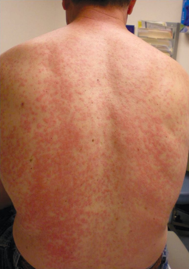
 A morbilliform drug eruption consists of symmetrically arranged erythematous macules and papules, some discrete and others confluent.
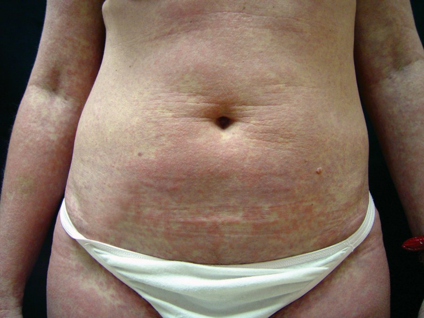
Urticarial reactions are the second most common type of cutaneous drug reaction and represent a type I hypersensitivity with IgE-mediated release of histamine. Wheals, pink-to-red effervescent edematous plaques with a surrounding flare, appear within minutes to a few hours after exposure to the causative agent (Figure 24). Patients may experience a spectrum of severity, from a few isolated wheals, to angioedema if the cutaneous edema becomes deep and confluent. In the most severe cases, anaphylaxis can occur, causing airway compromise and cardiovascular collapse. Nonimmunologic histamine release secondary to medications results in redness or flushing of the skin and variable pruritus without urticaria. Treatment of urticarial reactions is dependent on severity, with minor cases treated as described for exanthematous reactions, whereas anaphylaxis may require epinephrine, fluid resuscitation, and airway protection (for more information, see Urticaria).
 Urticarial drug reaction to amoxicillin with puffy pink plaques and an annular configuration.
Fixed drug reactions recur in the same site (or sites) on the skin with each exposure. Pink-to-purple circinate plaques with central dusky discoloration or vesiculation appear most commonly on the lips, face, fingers, and genitals (Figure 25). The first exposure typically generates one plaque that recurs in the same location with each repeat encounter. Additional plaques may develop with subsequent exposures. Lesions typically resolve with muddy brown postinflammatory hyperpigmentation.
Photosensitivity disorders are a collection of conditions that have in common an abnormal skin response following exposure to ultraviolet radiation. Conceptually, it is useful to consider photosensitivity disorders that are idiopathic and those that are associated with application or ingestion of a drug or agent that causes cellular damage when exposed to ultraviolet radiation. Photosensitivity reactions related to exogenous agents are either phototoxic or photoallergic.
Polymorphous light eruption is the most common idiopathic photosensitivity disorder. It typically manifests before the age of 30 and is most common in fair-skinned women, first appearing in the spring and early summer. The rash will persist for weeks and resolve without scarring even with continued exposure to the sun. Lesions appear within hours of sun exposure and are found on the sun-exposed body parts. While many different types of eruptions may occur, the most common are pruritic skin-colored or pink papules. Other variants include vesicles, bullae, and plaques. The type of lesion will remain consistent in an individual with each occurrence.
Phototoxicity can occur in anyone with a threshold concentration of the responsible chemical or drug and is more common than photoallergy. Photoallergy is a cell-mediated immune response found in previously sensitized individuals that can be elicited by small amounts of offending agent. Phototoxic reactions present as an exaggerated sunburn within minutes to hours of sun exposure. Photoallergic reactions occur more commonly after topical exposure of a sensitizing agent and present as pruritic, eczematous eruptions within 24 to 48 hours after sun exposure on sun-exposed body parts (Figure 26). Treatment of photosensitivity disorders includes avoidance of allergens and photosensitizing drugs and broad-spectrum sunscreens if testing has ruled out sunscreen sensitivity.
Drugs are the most common cause of hypersensitivity vasculitis, a small vessel vasculitis. The American College of Rheumatology proposed criteria for the diagnosis of hypersensitivity vasculitis in a patient with vasculitis that are moderately sensitive and specific. Suggestive findings include palpable purpura and/or petechiae, which may be associated with fever, urticaria, arthralgia, and lymphadenopathy. These findings are typically associated with a low complement level and elevated erythrocyte sedimentation rate. The mainstay of treatment is drug discontinuation and avoidance. Severe or persistent symptoms may require systemic glucocorticoids. Medications can also cause a small/medium vessel ANCA-associated vasculitis (see Cutaneous Manifestations of Systemic Disease).